Single Electrodes
(Single Core Fiber Microelectrodes)
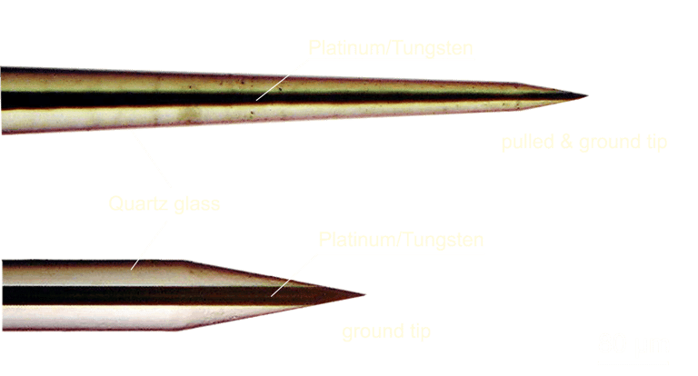
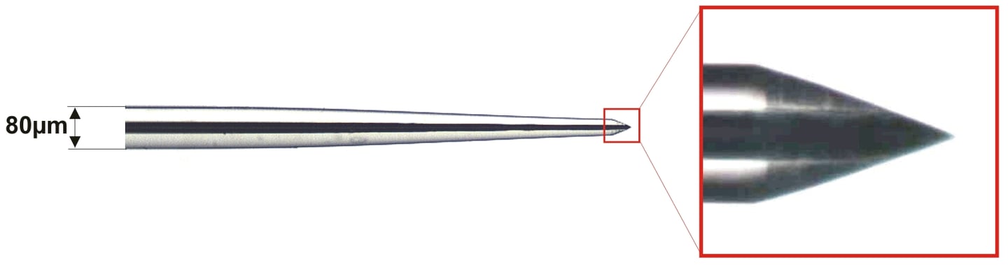
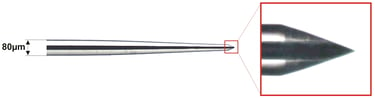
Thomas quartz/platinum-tungsten fiber microelectrodes permit the recording of extracellular potentials with excellent single-unit isolation, signal to noise-ratio and temporal stability. Thomas RECORDING GmbH offers a complete line of metal microelectrodes for recording extracellular potentials from single units and cell populations. The electrodes are very well suited to be applied to the electrode manipulators manufactured by Thomas RECORDING GmbH, but they may also be used for other applications. The electrode fibers are available with shank diameters ranging from 20 µm to 120 µm. Standard measures of our quartz-platinum/tungsten electrode fibers are 80 µm for the outer shank diameter and about 25 µm for the diameter of the metal core (other diameters on request!). A special developed manufacturing technique makes it possible, for tips to be drawn on these thin fibers, with both the outer quartz mantle and the metal core tapering down to very small dimensions. With a specific grinding technique the metal core is subsequently exposed to the required tip size respectively tip impedance (see “Manufacturing Equipment”).
Product Description

Tip Shapes
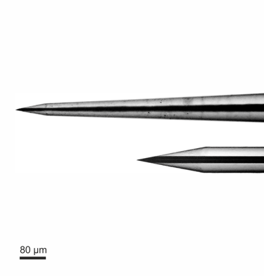
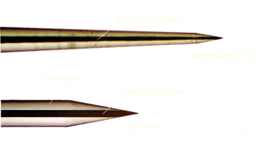
Our microelectrodes are available with two different tip profiles (see left picture).
Electrodes with ground tips (tip shape D) are very well suited for recording of multi-unit-activity (MUA), local-field-potentials (LFP) and for microstimulation of brain tissue (Impedance value at 1kHz in the range of 300kΩ to 800kΩ, depending on the fiber shank diameter and the grinding angle).
Electrodes with pulled and grinded tips (tip shape A), are very well suited for single-unit-recordings (Impedance value at 1kHz in the range of 1 MΩ to more than 10 MΩ in steps of 1MΩ, depending on the fiber shank diameter and the grinding angle.
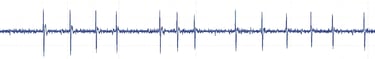
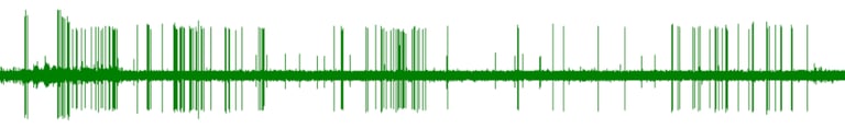
Recording Quality
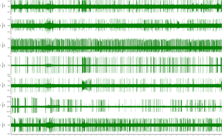
The picture on the rigth side shows an extracellular recording in primate amygdala with 7 Thomas quartzglass insultated platinum/tungsten fiber-microelectrodes loaded in a Thomas RECORDING multielectrode drive type 7 Electrode Eckhorn Matrix.
(with kind permission of Katalin M. Gothard, M.D., Ph.D. Associate Professor of Physiology and Neuroscience, Arizona Health Sciences Center, Tucson, Arizona, USA).
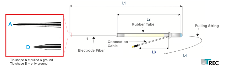
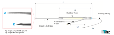
Fiber-microelectrodes
for TREC Rubber Tube Drives
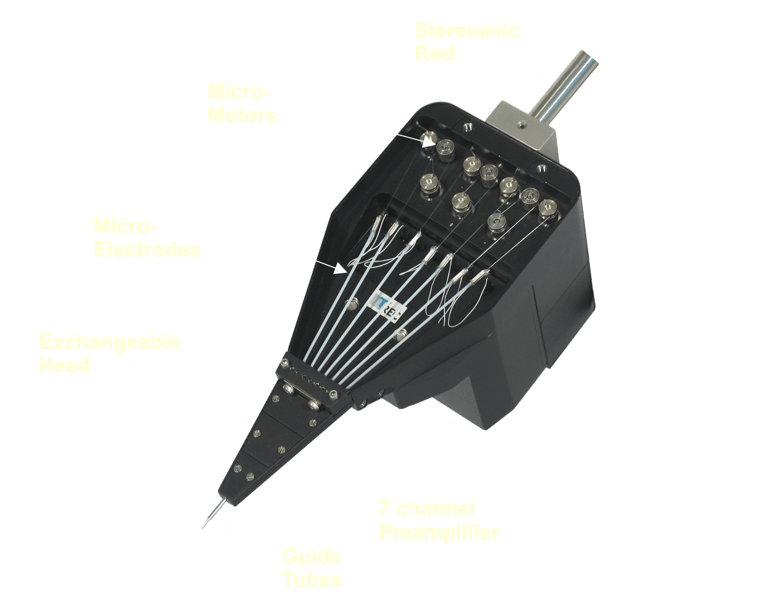

The fiber microelectrodes are available for Thomas RECRODING multielectrode manipulators with patented rubber tube drive. Left picture shows a TREC microdrive type "Eckhorn Matrix" loaded with 7 single core fiber electrodes.
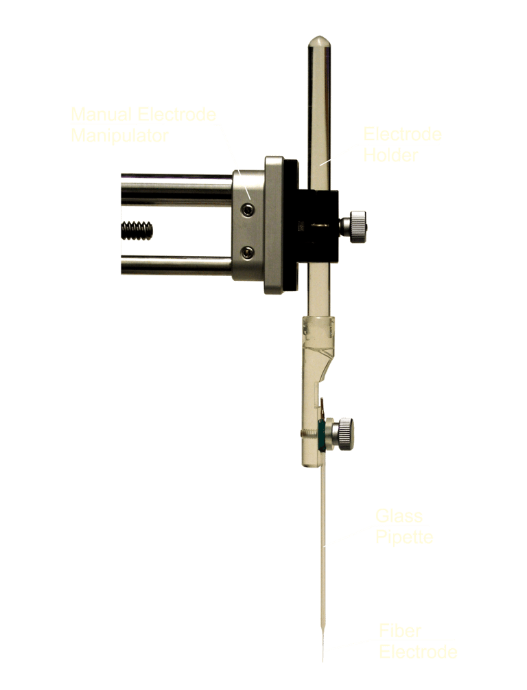
Fiber-microelectrodes
for other microdrives
The fiber microelectrodes are also available for standard manual melectrode manipulators or motorized single shaft electrode manipuators like the Thomas RECRODING MEM. The picture on the right side shows a TREC manual microdrive loaded with single core fiber microelectrode.
The electrode fiber is strengthened with a glass pipette so that the electrode can be clamped to the electrode holder.


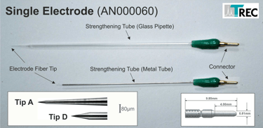
Article number: AN000060
Data Sheet
Article number:
AN000050 (Eckhorn Matrix, 1 piece)
AN000222 (Eckhorn Matrix, box of 12 pieces)
AN000203 (Mini Matrix, 1 piece)
AN000204 (Mini Matrix, box of 12 pieces)
AN000217 (Tetrode Mini Matrix, 1 piece)
AN000218 (Tetrode Mini Matrix, box of 12 pieces)
Data Sheet
Advantages of TREC Fiber Microelectrodes
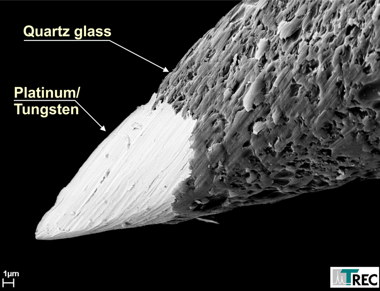
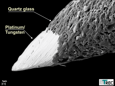
The passage between the glass isolation and the core of all of our microelectrodes is very smooth (see figure 1 below). The result is, that there will be not so much tissue damage during the electrode penetration. Other electrodes like silicon-substrate electrodes or isolated metal-wire electrodes usually have cutting edges or a terraced passage between the core isolation and the tip and they will cause more tissue damage. Due to their smooth shape and their small dimensions, our microelectrodes cause only minimal tissue damage. Tissue damage by our microelectrodes, in fact, is so small that electrode tracks cannot be verified with standard histological techniques!
Due to the geometrical shape of our fiber microelectrode tips, brain tissue is displaced radially during penetration, with little tissue compression. There is little or no-readjustment of tissue after insertion, which might be one of the reasons why injury potentials are hardly ever seen after electrode movement, has been stopped.
Microgrooves, caused by the grinding process, increases the effective tip area at a given tip volume (see figure 2 on the left side).
This results in a tip capacitance of more than 2pF/µm2, which is considerably higher than the tip capacitance of etched tips. This high tip capacitance of our microelectrodes is one reason for their excellent signal-to-noise ratio and single-unit isolation. So our quartz-platinum/tungsten fiber electrodes permit the recording of extracellular potentials with excellent single-unit-isolation, signal-to-noise ratio and temporal stability
The shaft diameter of the most conventional microelectrodes, generally, is relatively large (up to 300µm). It is therefore, not possible to use such electrodes in closely spaced parallel arrays for multi-channel recordings. The shaft of our microelectrodes, over the whole penetration depth, is cylindrical and very thin (see figure 3). Our standard microelectrode has a shaft diameter of 80µm. Smaller diameters down to 20µm are also available. So our fiber microelectrodes are well suited for multi-electrode arrays. In contrast to silicon-substrate electrodes (Wise and Angell, 1969; Wise and Starr, 1969) movement control of individual electrodes is possible.
Our microelectrodes have a wide recording bandwidth and a low cut-off-frequency, so that both, spike potentials and (slow) local filed potentials can be recorded from the same microelectrode.
The electrodes are strong enough to penetrate the intact dura of monkey or cat for a relatively long time. Since the dura does not to be opened, the preparation is simplified and danger of trauma and of infection in chronic preparations is greatly reduced.
If our electrodes are used together with Thomas Microdrive systems, it is possible to drive the electrodes over long distances through the brain (e.g. 40mm). The electrodes will move straight and do not leave the trajectory to the target (e.g. primate amygdala). See figure 3 below.
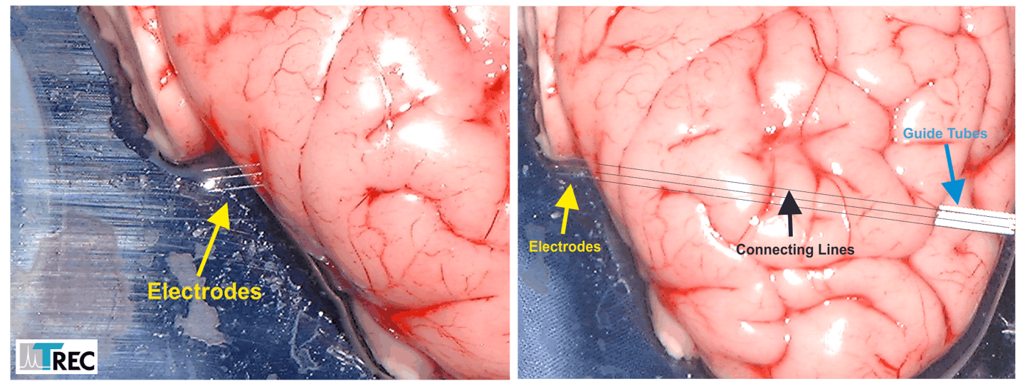
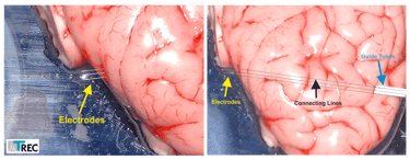
Figure 3: These pictures show a penetration test with Thomas fiber microelectrodes. The electrodes moves straight through the brain over a distance of 40.000µm although the fibers just had 80µm outer diameter. The drawn connecting lines in the rigth photo show that the exit points of the elelctrodes are exactly at the opposite side of the entrance points.
Figure 2: Microgrooves in the metal increase the effective metal area which causes a higher tip-tissue capacity, one reason for the excellent signal to noise ratio of these electrodes.
Because of our precision manufacturing techniques the geometrical shape of the electrode tip can be made exactly and reproducibility according to specifications, as long as it is a tip geometry that is grindable. Conical tips can be reproduced with high accuracy (see figure z). With our precision manufacturing equipment tip geometry of the microelectrodes can be optimized for a particular recording situation. Each microelectrode will be double checked, electronically and microscopically. We give the impedance value for each electrode, documented in a test certificate, enclosed to the electrode delivery.
We are able to produce microelectrodes that will meet the special requirements of our customers. For example different electrode lengths, different tip profiles, special impedance values (for single-unit isolation, multi-unit isolation, local field potential recordings or stimulation) are available. Please ask for your special configuration.
Electrode tip manufacturing equipment is also available from Thomas RECORDING GmbH. With this equipment the scientist will be able to manufacture his own microelectrodes for his special neurophysiological application.
Figure 1: The passage between the glass insulation and the metal part of the electrode tip is so smooth, that only minimal tissue damage is caused when the electrode is introduced in brain tissue.
Product Information
Marking of Recording Position
[1] Reitboeck, H.J.
Fiber microelectrodes for electrophysiological recordings,
J Neurosci Methods 8, 249-262 (1983).
[2] Reitboeck HJ.
A 19 channel matrix drive with individually controllable fiber microelectrodes for neurophysiological applications.
IEEE Transactions on Systems, Man and Cybernetics 1983; 13: 676-683.
[3] Eckhorn R, Thomas U.
Fasermikroelektroden für technische und medizinische Anwendungen.
Hannover Messe. 1987.
[4] Mountcastle V.B., Reitboeck H.J., Poggio G.F., Steinmetz M.A.
Adaptation of the Reitboeck method of multiple microelectrode recording to the neocortex of the waking monkey,
Journal of Neuroscience Methods, 36 (1991) 77-84
[5] Eckhorn R, Thomas U.
A new method for the insertion of multiple microprobes into neural and muscular tissue, including fiber electrodes, fine wires, needles and microsensors.
J Neurosci Methods 1993; 49: 175-179.
Selected Publications
Request Information
Location
Winchester Strasse 8
35394 Giessen, GERMANY
Hours
Mo.-Fr. 8:00 a.m - 4:00 p.m.
Central European Time (CET)
Sa. - Su. Closed
info@ThomasRECORDING.com