


Tip Types
The Thomas Optical Fiber is available with blunt tip as shown in figure 1 or with a conical tip like shown in figure 2.
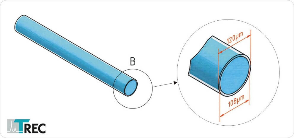 Figure 1: Thomas Optical Fiber dimensions. Core glass diameter: 108µm, outer shaft diameter: 120µm, Material: glass
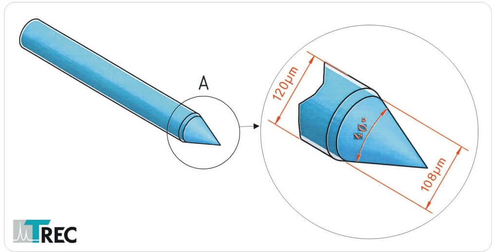 Figure 2: Thomas Optical Fiber conical tip
Tip Size
The Thomas Optical Fiber with conical tip is available in three different shapes like shown in figure 3.
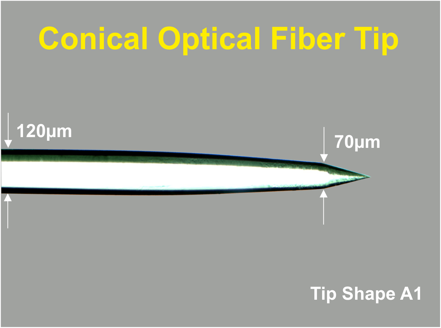 Figure 3A: Thomas Optical Fiber with double conical (Tip A1) tip shape
 Figure 3B: Thomas Optical Fiber with double conical (Tip A2) tip shape
 Figure 3C: Thomas Optical Fiber with conical (Tip D)
Advantages of Thomas Optical Fibers
The advantage of Thomas conical and double conical fibers is that they do not cause tissue damage when introduced in brain tissue in comparison to blunt glass fiber tips. Furthermore Thomas Optical Fibers have a globular light distribution at the conical tip (see figure 4) in contrast to the cylindrical light distribution of blunt glass fibers.
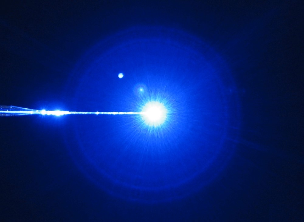 Figure 4: Globular light distribution of a Thomas Optical Fiber with conical tip shape
Versatile
Thomas Optical Fibers are available for use in Thomas Microdrive systems (e.g. Mini Matrix, Eckhorn Matrix, see figure 5) and also for use with other manipulators (see figure 6).
 Figure 5: A single Thomas optical fiber and 6 Thomas microelectrodes loaded to a 7 channel microelectrode manipulator system “Eckhorn Matrix”. The Microdrive is equipped with a special head allowing a concentric arrangement of electrodes and optical fiber. Each electrode and optical fiber are independently moveable to different depths of the brain. Cortical as well as deep brain stimulation/recording experiments are possible. The lateral distance between electrodes and optical fiber is 254µm but can be reduced down to 80µm!
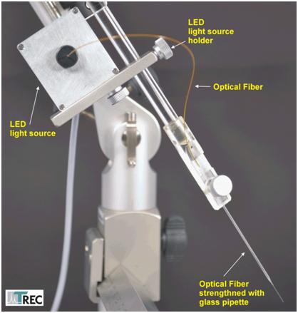 Figure 6: Thomas Optical Fiber mounted to a small animal stereotaxic instrument electrode holder

NEWS

PRODUCTS

SOLUTIONS

DISTRIBUTORS
 Sign Up