
Overview
With this fiber-electrode manipulator type Mini Matrix a broad range of even very thin shafted probes can be handled, including fiber and wire-electrodes with shaft diameters down to about 25 µm. The patented driving principle of the Mini Matrix offers an outstanding electrode positioning accuracy. Compared with hydraulic, cable controlled or direct motor driven microdrives, the patented fiber electrode manipulator “System Eckhorn” does not cause hysteresis errors of the electrode movement. Hysteresis error is generally a result of stiction and free motion of hydraulic, cable controlled or direct motor driven microdrives offered by our competitors. Our system has a higher degree of positioning accuracy due to its patented rubber tube driving mechanism being almost absolutely free of stiction and free motion.
Using Thomas Mini Matrix systems with patented rubber tube drive allows to record neural activity while the electrode is moving in the brain. So it is very easy to search active neurons in the brain.
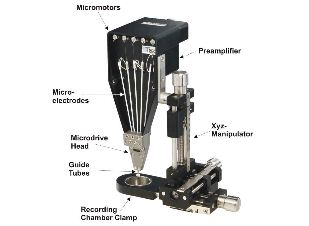 Figure 1: System components of a 5 Electrode Mini Matrix for primate recording applications. The 5 electrode Mini Matrix is equipped with a xyz-manipulator which has a primate recording chamber clamp. We can adapt the chamber clamp to the outer diameter of the recording chamber.
The Thomas Mini Matrix systems have some advantages in comparison to other multielectrode microdrive systems for neurophysiological research presently available on the market:
- Vibration-free positioning of up to 5 quartz-platinum/tungsten microelectrodes in 1µm steps
- Patented rubber tube driving principle
- High positioning accuracy, due to lack of stiction and free motion
- Easy exchange and calibration of microelectrodes within minutes
- Advances up to 5 microelectrodes/tetrodes independently in 1µm steps up to 24.000µm (other electrode travel distances on request!)
- Xyz-manipulator travel-distance: z=0-30mm, x= ±10mm, y=±10mm
- Continuous low noise recording while the electrode scans the tissue at low velocity
- No noise introduction in the recordings so that you can search active neurons in the brain
- No electrode connection cables free in air, no electrical noise pickup from environment (e.g. motors, radio stations)! Competitor drives pickup electrical interference from the environment so that you for example can hear radio station music or electrical artifacts from lab devices from the audio monitor loudspeaker instead of neural spikes!
- Electrode moving velocity adjustable (1-250µm/s), moving direction selectable
- Microdrive with xyz-manipulator is small and lightweight
- Low noise 5 channel preamplifier is integrated into the microdrive chassis, system is completely shielded to avoid electrical noise pickup from the environment, three preamplifier operation modes: record, electrode impedance test, electrical stimulation/lesioning
- System is completely delivered with motor control device, integrated preamplifier, software, xyz-manipulator, chamber holder, exchangeable head, handheld remote control, etc.
- Exchangeable manipulator head allows manifold electrode configurations and interelectrode spacings
- Electrode spacings from 80µm up to some millimeters possible due to exchangeable Mini Matrix heads
- The system is modular and adaptable to the end user´s requirements.
- The Mini Matrix is usable for cortical as well as deep brain recordings in all kinds of research applications
- User friendly computer controlled system with LAN (local area network) communication via TCP/IP protocol between motor control unit and personal computer. Software package is part of the system.
- Using very thin shaft Thomas microelectrodes (outer diameter 80µm) causes minimal tissue damage.
- Different neurophysiological methods are available for the Mini Matrix systems (e.g. optogenetics, microinjection)
The Mini Matrix system is available for small animal stereotaxic instruments (see figure 3 and 4) as well as for primate experiments (see figure 2).
 Figure 2: 5 channel electrode Mini Matrix mounted on a primate recording chamber.
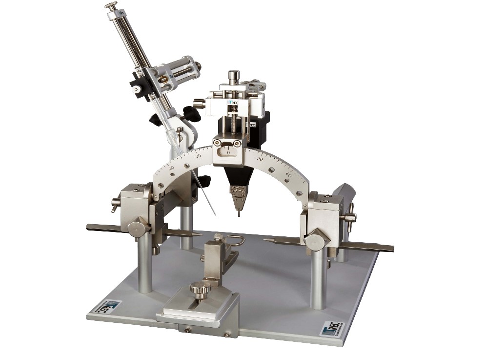 Figure 3: Mini Matrix mounted to a Thomas small animal stereotaxic instrument (SASI). By using customized adapters the Mini Matrix can be mounted on all other stereotaxic instruments presently available on the market. Please feel free to ask you your special adaptation.

Exchangeable Heads
Each Mini Matrix microdrive is equipped with an exchangeable Microdrive head. Each Mini Matrix head has up to 5 electrode guide tubes. There are different possibilities to arrange these 5 guide tubes, depending on the recording area. Basically we have exchangeable heads for cortical, deep brain or spinal cord recordings.
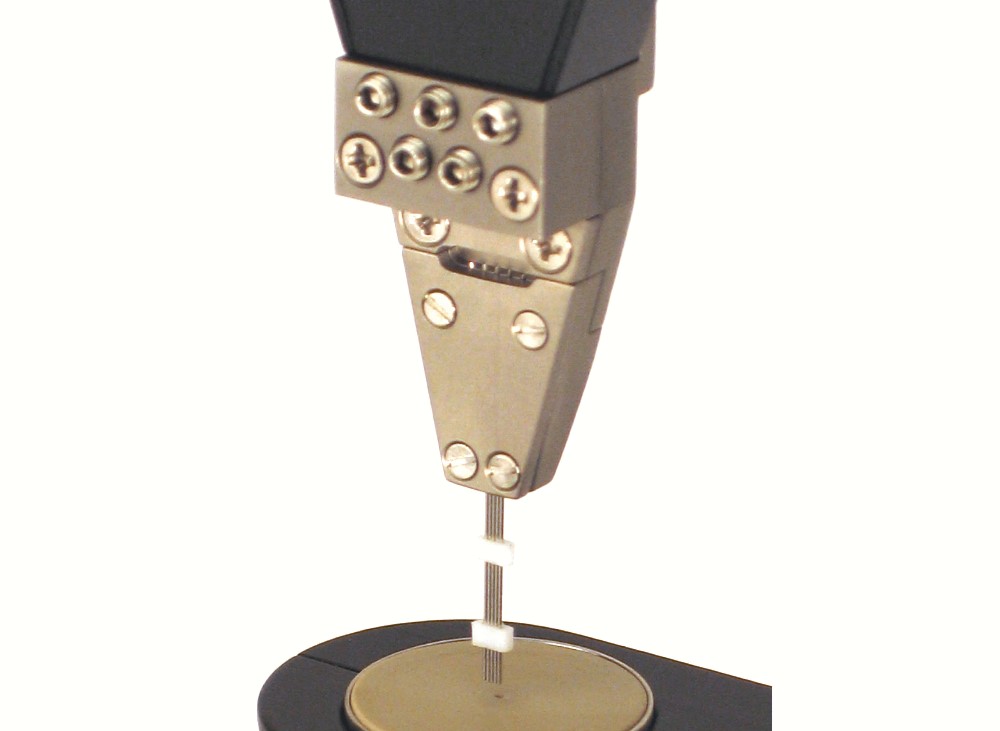 Figure 5: 5 channel Mini Matrix head with linear electrode configuration and 305µm electrode spacing.
It is very easy to exchange a Mini Matrix head by another one with other electrode guide tube arrangement or electrode guide tube spacing. The following picture demonstrates the exchange procedure.
 Figure 6: Exchange of Mini Matrix head
Heads for cortical recordings
There are four parameters of an exchangeable Mini Matrix head that can be customized:
- protrusion of the electrode guide tubes
- arrangement of the guide tube (e.g. linear, concentric, etc.)
- guide tube spacing (e.g. 305µm, 500µm. etc.)
- Sharpened of non-sharpened (standard) guide tubes for dura penetration
The standard guide tube protrusion for an exchangeable head for cortical recordings is x=8mm, which is optimal for most recording applications (e.g. rats, mice, rodents, etc.). If you are using deep primate recording chambers we can adapt the guide tube protrusion to the depth of the primate recording chamber.
 Figure 7: Mini Matrix head with linear guide tube arrangement and electrode spacing of 305µm (other guide tube arrangements e.g. concentric and other spacing e.g. 500µm)
Heads for deep brain recordings
For deep brain recording applications in small animals the normal electrode travel distance of a Mini Matrix system of 22-24 mm is enough to reach the deep brain target in rats, without introducing a cannula in the brain. For larger animals such as primates the use of a common guiding cannula with minimized outer diameter is required to bring up to 5 microelectrodes down to the recording target. Therefore we offer a special cannula technique for our Mini Matrix, invented by Dr. David Leopold (NIMH, USA). We call this technique the “Leopold Funnel Adapter”.
 Figure 8: Mini Matrix head (5 channels) without (left side) and with (right side) Leopold funnel cannula. The funnel cannula guides all 5 microelectrodes together in one cannula. The electrode spacing is 80µm for single core electrodes and 100µm for tetrodes.
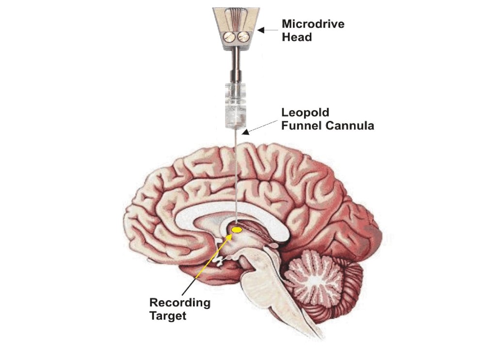 Figure 9: The funnel cannula is introduced in the brain down to app. 5mm before recording target under stereotaxic control. Then one drives the microelectrodes out of the cannula into the target.
Please feel free to ask for your customized deep brain recording solution!
Heads for spinal cord recordings
Microdrive heads for spinal cord recording have a linear guide tube arrangement and interelectrode spacings from 305µm up to 10mm, depending on the researcher´s requirements.
 Figure 10: Exchangeable Mini Matrix head for spinal cord recordings. The electrode guide tube arrangement is linear and the equidistant guide tube spacing is 5mm. Other spacing (smaller or larger) is available on request.
Electrical Stimulation and Impedance Test
The MSD is equipped with a special low current impedance test unit that allows measuring the electrode tissue impedance of each microelectrode while the electrode is in the brain. The constant current is so low (5nA) that it does not stimulate nerve cells in the brain. The electrode impedance value is displayed on a moving coil instrument. We have used this kind of instrument instead of digital because moving coil instruments better display fluctuations in the impedance value which is important especially when electrodes penetrate tissue borders (e.g. primate dura mater).
 Figure 11: Mode Selection & Impedance Device consists of a (1) control box and a (2) main device which can be mounted in a 19” rack or on a workbench.
 Figure 12:MSD for 5 Tetrode Mini Matrix systems. The impedance test module with the moving coil instrument is located on the right side of the front panel.
The preamplifiers of our Mini Matrix systems are prepared for electrical stimulation and lesioning. You can pass a stimulus or lesioning current through each of the 5 microelectrodes. A Mode Selection & Impedance Test Device (MSD, see figure 12) is required as well as a microstimulation generator. Both devices are available from TREC on request.
Especially for microstimulation we offer special coated Thomas microelectrodes. The microstimulation electrode tips are coated with iridium oxide. This coating increases the charge transfer capacity of our electrodes dramatically which makes them very well suited for stimulation experiments.
Setting lesions with Thomas microelectrodes
At the end of a recording session you might want to mark the recording position of the recording electrode. In this case you can pass a lesion current through our standard microelectrode tip. It could be shown by Robert Shapley and colleagues, that small dc currents make good lesions in primate visual cortex without any electrode tip damage (see unpublished report).
Microinjection with the Mini Matrix
For making drug injection experiments with the Thomas Mini Matrix system we offer a special designed high pressure microinjection system. One of the 5 single microelectrodes of the Mini Matrix can be replaced by a microinjection cannula. The microinjection cannula (OD=120µm) has a tapered tip and is equipped with the patented Thomas rubber tube drive so that you can position the microinjection cannula tip together with the recording microelectrodes with high positioning accuracy (better than 1µm). The microinjection system consists of a special designed and computer controlled syringe pump that is connected to the microinjection cannula via a special thick wall tubing.
The precision micromotor of the microinjection pump is controlled by the same motor control unit and graphical user interface as the Thomas Mini Matrix. This guarantees a high precision and convenient experimental control of the injection/recording experiment.
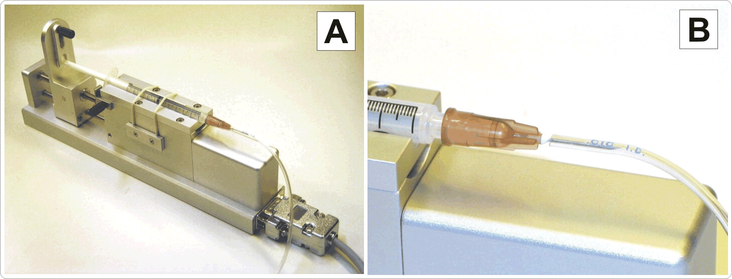 Figure 13: Microinjection pump (A), connection of thick wall tubing to syringe cannula (B)
Optogenetic equipment for the Mini Matrix
Thomas RECORDING offers a complete range of Mini Matrix accessories for optogenetic experiments. One of the 5 single microelectrodes or tetrodes is replaced by an optical fiber (OD=120µm). The optical fiber has a tapered tip and is equipped with the patented Thomas rubber tube drive so that you can position the optical fiber tip together with the recording microelectrodes with high positioning accuracy (better than 1µm). Figure 14 shows a Mini Matrix head with concentric guide tube arrangement and a single optical fiber in the center position. The recording microelectrodes are concentrically arrange around the optical fiber in the center of this arrangement.
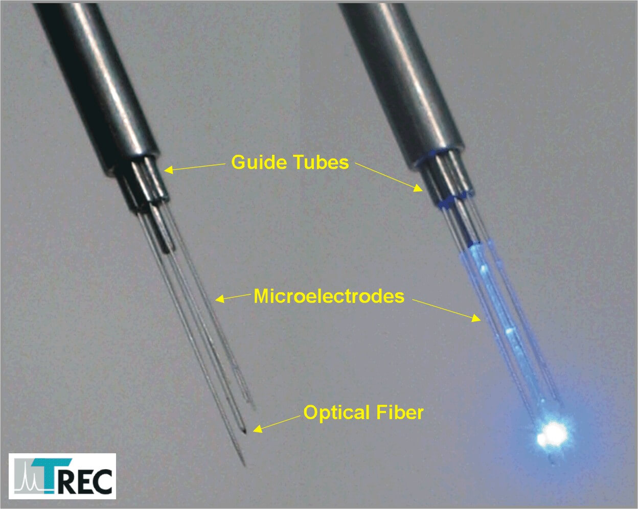 Figure 14: Mini Matrix concentric head with one optical fiber and 4 recording electrodes.
In figure 15 one can see a Thomas Mini Matrix equipped with a LED-light source holder. In figure 16 an optical fiber is loaded to the Mini Matrix.
 Figure 15: Thomas Mini Matrix with xyz-manipulator, LED light source and light source holder
 Figure 16: Thomas Mini Matrix with LED light source and loaded optical fiber.
For further information concerning optogenetic equipment please see also our optogenetic webpage.

NEWS

PRODUCTS

SOLUTIONS

DISTRIBUTORS
 Sign Up