THOMAS RECORDING

Home   >   Products   >  Neuroscience Products   >  Screws   >  Ceramic Screws   >   Ceramic Screws

Neuro. Prod. > Accessories > Amplifiers > Chron. Rec. Dev. > Data Acquis. Sys. > Eye Track. Sys. > Microdrives > Microelectrodes > Microinject. Sys.> Optogenet. Prod. > Prim. Train. Sys. > Screws > Software > Stereotaxic Instr. > Cell&Tissue Med. > Rodent Products Medical Products Electrochemical Products

Ceramic Screws

MRI- and bio-compatible screws

Neuroscience Products > Accessories > Amplifiers > Chronic Recording Devices > Data Acquisition Systems > Eye Tracking Systems > Microdrives > Microelectrodes > Microinjection Systems> Optogenetic Products > Primate Training Systems > Screws > Software > Stereotaxic Instruments > Cell & Tissue Media > Rodent Products Medical Products Electrochemical Products

Ceramic Screws

MRI- and bio-compatible screws

For MRI applications care must be taken to limit the amount of magnetically susceptible metal inside the MRI scanner. Particularly, if any screws are included for implant fixation, the use of ceramic material will limit the distortion caused to the MRI scan.

Key features:

  • MRI-compatible screws
  • Material: ceramic
  • Two differend screw types
  • SI screws
  • SA screws
  • Tools for the screws available
Technical Data
Features
Downloads
Data 
Material:Ceramic
Dimensions:Variable, please see figures for available types of ceramic screws

Features

Therefore it is recommended to fix implants with ceramic screws. Thomas RECORDING offers a set of 5 different ceramic screws made of zirconium oxide.

These screws were developed and firstly used by Prof. Dr. Nikos Logothetis at the Max Planck Institute for Biological Cybernetics in Tuebingen, Germany [1]. The ceramic screws were tested on MR quality control phantoms and were found to have no effects on the homogeneity of the B0 field of the magnet [1]. In addition the material was chosen to be tissue compatible and are surface roughened to optimize the bone and skin implant interface [1].

Thomas RECORDING is proud to be the worldwide sole distributor of these high quality ceramic screws. Our ceramic screws are used by leading neuroscientists all over the world.

Ceramic Screw Type SI

The ceramic screw type SI was the first version available from Thomas RECORDING for neurophysiological applications. It is a hexagon socket countersunk head screw available in two different total lengths of 6.4mm and 8.4mm (see figure 2 and 3). The cylindrically portion of the screw from the underside of the head to the tip is known as the shank which is partially threaded. The distance between each thread is called “pitch”. The thread of the SI screws looks like the one of self-cutting titanium bone screws but it is different. The ceramic screw is not self-cutting. Therefore a special drill and wrench is required to insert these screws in bone tissue (see TREC ceramic screw tools). These screws are not intended to be used in human medical applications.

Figure 2: Ceramic screw SI04 (AN000053)

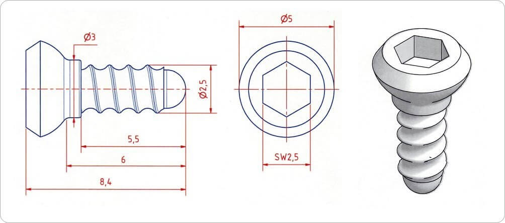
Figure 3: Ceramic screw SI06 (AN000054)

Ceramic Screw Type SA

The ceramic screw type SA is a later development. It is a hexagonal socket (hex socket, Allen) screw available in five different total lengths of 5.9, 8.0, 9.2, 11.1 and 13.1mm (see figures 4 to 8). The cylindrically portion of the screw from the underside of the hexagon socket head to the tip is known as the shank which is partially threaded. The distance between each thread is called “pitch”. The thread of the SA screws also looks like the one of self-cutting titanium bone screws but it is different. The ceramic screw is not self-cutting. Therefore a special drill and wrench is required to insert these screws in bone tissue (see TREC ceramic screw tools).

Figure 4: Ceramic screw SA05 (AN000056)

Figure 5: Ceramic screw SA06 (AN000057)

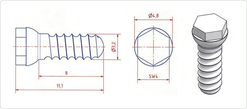
Figure 6: Ceramic screw SA08 (AN000058)

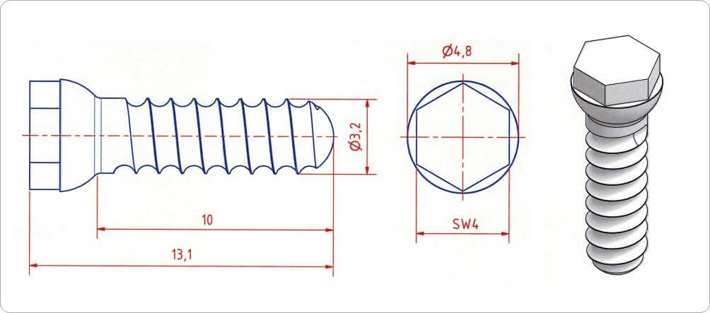
Figure 7: Ceramic screw SA10 (AN000059)

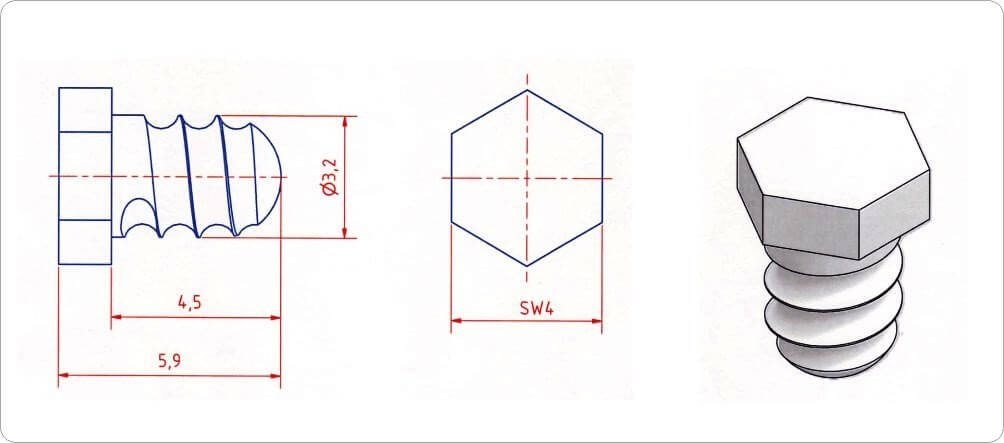
Figure 8: Ceramic screw SA45 (AN000055)

Publications

[13] Dougherty, K., Carlson, B. M., Cox, M. A., Westerberg, J. A., Zinke, W., Schmid, M. C., … & Maier, A. (2021). Binocular suppression in the macaque lateral geniculate nucleus reveals early competitive interactions between the eyes. Eneuro.

[12] Lee S.-K., Hwang S.-H., Barg J.-S., Yeo S,.J. Rapid, theoretically artifact‐free calculation of static magnetic field induced by voxelated susceptibility distribution in an arbitrary volume of interest. March 2018, DOI: 10.1002/mrm.27161

[11] Srivatsun Sadagopan, Wilbert Zarco, Winrich A Freiwald, A causal relationship between face-patch activity and face-detection behavior. eLIFE Sciences, April 2017, DOI: 10.7554/eLife.18558

[10] Karolina Marciniak, Artin Atabaki, Peter W Dicke, Peter Thier, Disparate substrates for head gaze following and face perception in the monkey superior temporal sulcus. eLIFE Sciences, July 2014

[9] Melanie Wilke, Igor Kagan, Richard A. Andersen, Functional Imaging Reveals Rapid Reorganization of Cortical Activity after Parietal Inactivation in Monkeys. Proceedings of the National Academy of Sciences of the United States of America ( PNAS ), May 2012 (please see  supporting information)

[8] Joseph Feingold, Theresa M. Desrochers, Naotaka Fujii, Ray Harlan, Patrick L. Tierney, Hideki Shimazu, Ken-ichi Amemori, Ann M. Graybiel, A system for recording neural activity chronically and simultaneously from multiple cortical and subcortical regions in nonhuman primates. Journal of Neurophysiology, December 2011

[7] Mark S. Bolding, Meredith A. Reid, Kathy B. Avsar, Rosalinda C. Roberts, Paul D. Gamlin, Timothy J. Gawne, David M. White, Jan A. den Hollander, Adrienne C. Lahti, Magnetic Transfer Contrast Accurately Localizes Substantia Nigra Confirmed by Histology. Biological Psychology, July 2012

[6] Anil Bollimunta and Jochen Ditterich, Local Computation of Decision-Relevant Net Sensory Evidence in Parietal Cortex. Cerebral Cortex, April 2012

[5] Elias B. Issa, James J. DiCarlo, Precedence of the Eye Region in Neural Processing of Faces. Journal of Neuroscience, November 2012

[4] Alexander Maier, Christopher J. Aura, David A. Leopold, Infragranular Sources of Sustained Local Field Potential Responses in Macaque Primary Visual Cortex. Journal of Neuroscience, February 2011

[3] Alexander Maier, Geoffrey K. Adams, Christopher Aura, David A. Leopold, Distinct superficial and deep laminar domains of activity in the visual cortex during rest and stimulation. frontiers in Systems Neuroscience, August 2010

[2] Scherberger H., Fineman I., Musallam S., Dubowitz D.J., Bernheim K.A., Pesaran B., Corneil B.D., Gilliken B., Andersen R.A., Magnetic resonance image-guided implantation of chronic recording electrodes in the macaque intraparietal sulcus. Journal of Neuroscience Methods 130 (2003) 1-8

[1]  Logothetis N., Guggenberger H., Peled S., Pauls J., Functional imaging of the monkey brain. nature neuroscience, vol. 2 no. 6, June 1999

Questions? Please don’t hesitate contacting us!

Data Privacy

* Required field

NEWS

PRODUCTS

SOLUTIONS

DISTRIBUTORS


SIGN UP TO OUR NEWSLETTER

Sign Up

Contact       Imprint       Data Privacy       Solutions       About Us       YouTube       Twitter